
UNDERSTANDING DENTAL X-RAYS:
Safety and Purpose

“Hey Dr. Tudo, are X-rays harmful? I don’t want to be exposed to unnecessary radiation.” As a dentist at Clarence Street Dental Group, I hear this question regularly — and I’m glad patients ask it. The honest answer is no, dental X-rays are not harmful when used appropriately. Modern dental imaging is among the safest in all of medicine, and the clinical information it provides is essential to delivering thorough, high-quality care.
X-RAY SAFETY
The amount of radiation from dental imaging is extremely small — comparable to the natural background radiation we are all exposed to every day. A single dental X-ray delivers roughly the same radiation dose as eating a few bananas. A short airplane flight or a day in the sun exposes you to more.
Dental imaging technology has also improved dramatically over the years. While older film-based X-rays required higher radiation doses, today’s digital sensors use up to 90% less radiation while producing clearer, more detailed images. This allows us to detect problems earlier, often before they become more serious or difficult to treat.
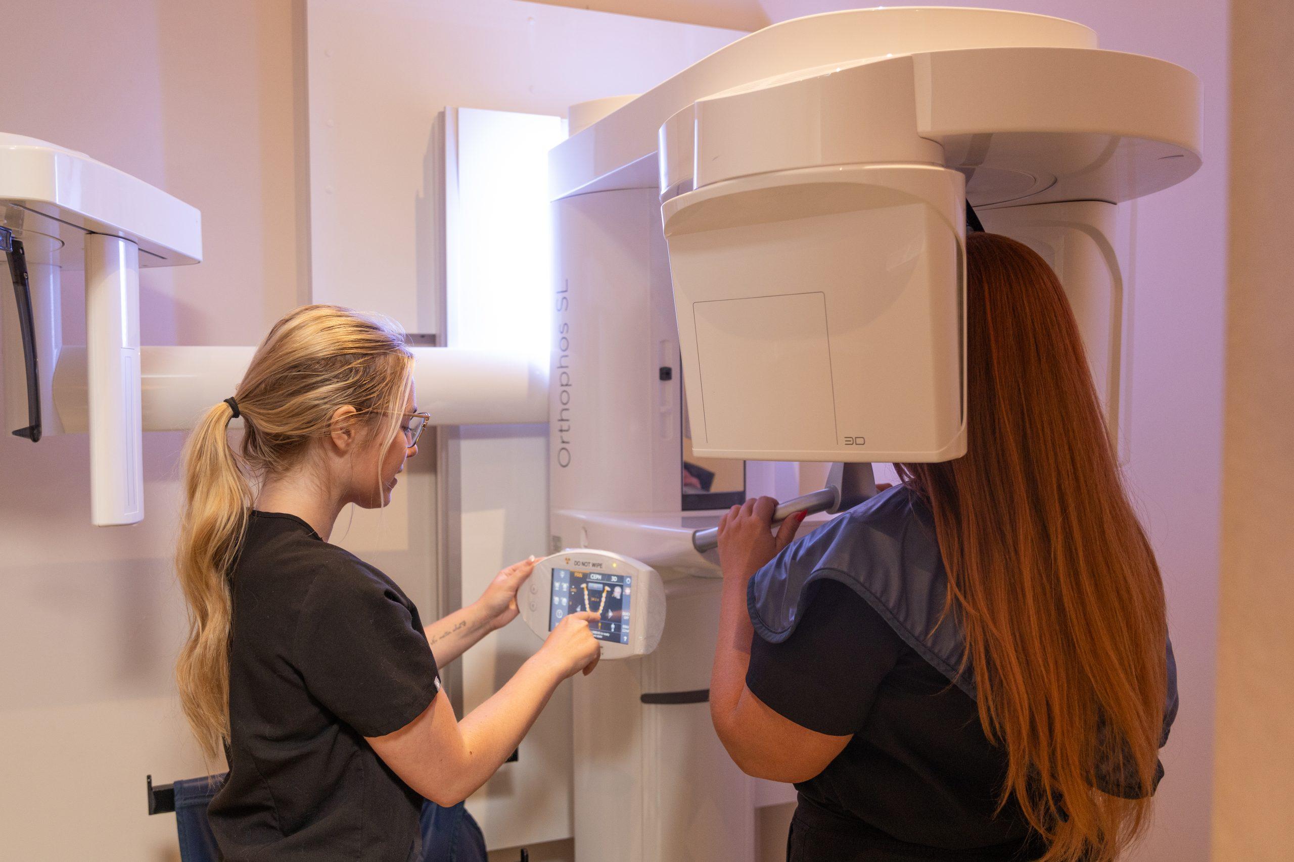
Regardless of the imaging type, your safety is always our first consideration. We follow the ALARA principle — keeping radiation exposure As Low As Reasonably Achievable — and always use lead aprons and thyroid collars. Radiographs are only recommended when there is a genuine clinical need; they are never taken out of habit or routine without a clear reason.
THE PURPOSE OF DENTAL X-RAYS
We don’t take a one-size-fits-all approach to dental imaging. Different clinical situations call for different types of radiographs, each chosen for a specific purpose:
Periapical X-rays — These allow us to examine individual teeth from crown to root tip. We use them to evaluate root health, look for signs of infection at the root apex, and assess the level of surrounding bone. They are essential for diagnosing abscesses, root fractures, and bone loss related to periodontal disease.
Bitewing X-rays — Our primary tool for detecting cavities (dental caries), particularly those forming between teeth where a visual exam cannot reach. They also reveal bone height between teeth. We take these at recall appointments to catch decay as early as possible, when treatment is most conservative.
Panoramic X-rays — These capture a broad, single view of the entire mouth — all the teeth, both jaws, the temporomandibular joints (TMJ), and surrounding structures. We use panoramic imaging to evaluate wisdom tooth development and position, screen for pathology such as cysts or tumors, and get an overall picture of jaw health that individual X-rays alone cannot provide.
Cephalometric X-rays — These produce a precise lateral (side) view of the skull and facial structures, used primarily for orthodontic treatment planning. They allow us to analyze the relationship between the teeth, jaws, and facial profile to guide decisions around braces, aligners, or corrective jaw surgery.
Cone Beam CT (CBCT) — This represents the most advanced level of dental radiographic imaging available, and one we are proud to offer at our practice. CBCT produces highly detailed, three-dimensional images of the teeth, bone, nerves, and surrounding anatomy. We use it for precise implant positioning, evaluation of complex pathology, planning conservative endodontic procedures, and accurately locating impacted teeth prior to surgical removal.
An important distinction: dental CBCT is not the same as the multi-slice CT scans used in hospital settings. The radiation dose from a dental CBCT is significantly lower than a hospital CT, while the level of anatomical detail and accuracy for dental and maxillofacial structures is superior.
Every X-ray we take at Clarence Street Dental Group has a purpose: to give us the clearest possible picture of your oral health so we can find problems early, plan treatment precisely, and help you maintain a healthy smile for life.